Shear Wave vs. Vibration-Controlled Elastography: What's the Difference?
Shear Wave vs. Vibration-Controlled Elastography: What’s the Difference?
When your doctor recommends a FibroScan to assess your liver health, you might not realize there are different technologies powering this non-invasive test. At Gastro Doctor, we specifically use Shear Wave Elastography, considered the more advanced evolution of liver stiffness measurement. But what sets it apart from the earlier Vibration-Controlled Transient Elastography (VCTE) technology?
Understanding this difference matters—not just technically, but for what it means for the accuracy of your diagnosis, especially if you’re being tested for immigration requirements or managing a chronic liver condition.
The Common Goal: Measuring Liver Stiffness
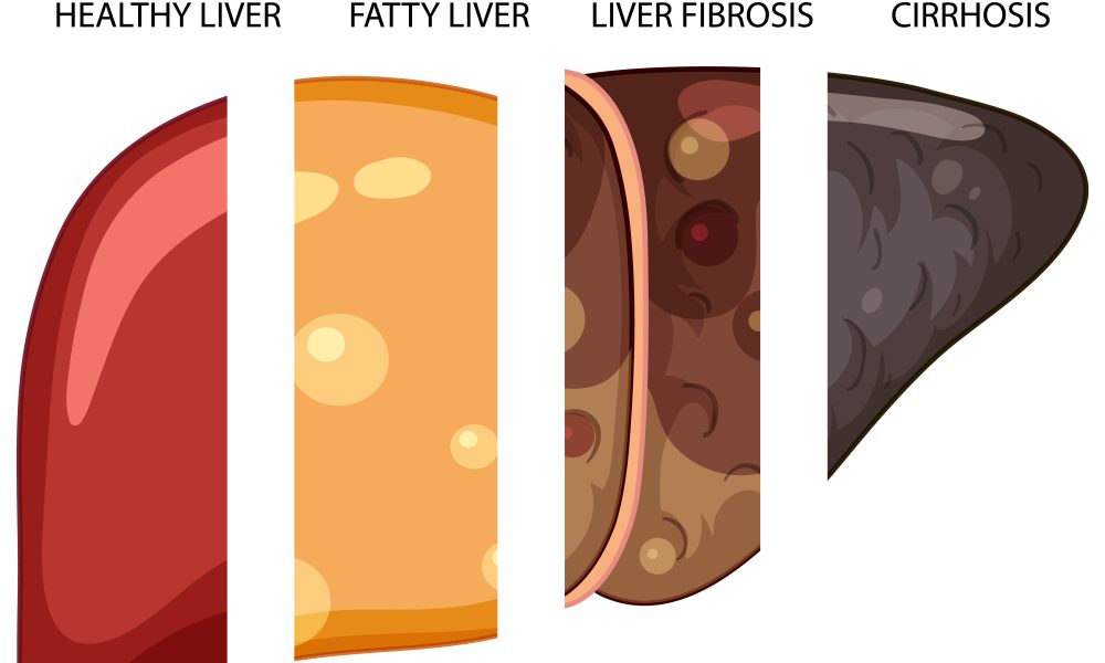
First, let’s establish what both technologies achieve brilliantly: they painlessly and safely measure liver stiffness, which correlates directly with the amount of fibrosis (scarring) in your liver. Both have rendered the invasive liver biopsy unnecessary for routine staging in most cases. This is a monumental win for patients.
The core difference lies in how they generate the measurement.
Technology Breakdown: Two Paths to a Measurement
1. Vibration-Controlled Transient Elastography (VCTE)
- The Original Technology: This is the method used in the very first generation of Shear Wave Elastography devices.
- How It Works: A probe placed on your skin generates a single, low-frequency mechanical vibration (like a tiny tap). This vibration creates a shear wave that travels through the liver. The device measures the speed of this wave—the stiffer the liver, the faster the wave travels.
- The “Transient” Part: It measures one wave per measurement, taking multiple measurements to get an average.
- Key Limitation: It’s often called a “blind” measurement. The probe doesn’t use real-time imaging to see exactly where in the liver it’s measuring. It assumes the tissue in its path is homogeneous liver tissue.
2. Shear Wave Elastography (SWE)
- The Advanced Technology: This is the method we use at Gastro Doctor. It represents the next generation, integrating ultrasound imaging.
- How It Works: Instead of an external mechanical tap, the ultrasound probe itself generates focused ultrasound “push pulses” within the liver tissue. These pulses internally create shear waves that fan out. The ultrasound then images these waves in real-time as they propagate.
- The “Imaging” Advantage: This happens while you’re looking at a standard ultrasound image of the liver on the screen. The operator can see exactly where they are measuring, avoiding large blood vessels, gallbladders, or nodules that could skew results.
- The Result: A shear wave velocity map (often a colour overlay on the ultrasound image) showing stiffness across a specific, targeted area.
Head-to-Head Comparison: Why the Difference Matters for You
| Feature | Shear Wave Elastography (SWE) | Vibration-Controlled (VCTE) |
|---|---|---|
| Core Technology | Ultrasound-generated “push pulses” inside tissue | External mechanical vibration |
| Real-time Imaging | ✅ YES. Uses standard B-mode ultrasound to guide and visualize. | ❌ NO. “Blind” measurement without imaging. |
| Targeting | Operator can select a specific, representative region of interest. | Measures a cylindrical volume of tissue along the probe axis. |
| Body Habitus | More reliable in patients with obesity. Can adjust depth and avoid thick subcutaneous fat. | Higher failure/error rates in obesity due to signal attenuation through fat. |
| Data Provided | Can simultaneously provide Controlled Attenuation Parameter (CAP) for fat quantification. | Provides stiffness (kPa) and separate CAP for fat. |
| Measurement Area | Smaller, targeted box (e.g., 1×2 cm) that can be placed precisely. | Larger, fixed cylindrical volume (1cm wide x 4cm long). |
| Accuracy Control | Visual confirmation that measurement is from valid liver tissue. | Relies on internal quality metrics (IQR/Median) but no visual backup. |
The Clinical Implications: Which is “Better”?
For most patients, both technologies will provide a reliable result. However, Shear Wave Elastography solves several key limitations, making it the preferred choice in specialist practice:
1. Superior Accuracy in Challenging Cases: For patients with a higher BMI or significant abdominal fat, VCTE can fail or give unreliable results. SWE’s ability to visualize and target deeper liver tissue makes it far more robust.
2. Avoiding Sampling Errors: The liver is not uniform. A VCTE measurement might inadvertently include a large blood vessel or a focal lesion, affecting the average. With SWE, the specialist can visually avoid these areas, ensuring the measurement is pure liver parenchyma.
3. Precision for Monitoring: When tracking subtle changes in liver stiffness over time (e.g., before and after starting treatment), the ability to measure the exact same region of the liver is a theoretical advantage of the targeting possible with SWE.
4. Confidence in Immigration Reporting: When providing a formal report for Immigration NZ, the robustness and visual verification of SWE add an extra layer of diagnostic certainty. There is a clear record (the ultrasound image with the measurement box) of what was measured.
Think of it this way: VCTE is like listening to a symphony to gauge the audience’s reaction. SWE is like having a high-resolution camera and a microphone, allowing you to see and hear the reaction of a specific section of the audience.
Why Gastro Doctor Invested in Shear Wave Technology
Our commitment, led by Dr. Dinesh Lal, is to provide the highest standard of diagnostic accuracy. Choosing Shear Wave Elastography reflects this principle:
- Evidence-Based: It is supported by a growing body of clinical literature as a highly accurate and reproducible method.
- Patient-Centered: It minimizes test failures and the need for repeat scans, reducing patient anxiety and inconvenience.
- Future-Focused: It represents the current technological standard in quantitative elastography, ensuring our patients benefit from the latest advancements.
The Bottom Line for Patients
If you are offered a Shear Wave Elastography, it’s a fantastic step for your liver health, regardless of the underlying technology. The most important factor is the expertise of the operator and interpreter—the specialist who performs the scan and puts the result into clinical context.
However, if you have the choice, Shear Wave Elastography offers distinct technical advantages in accuracy, reliability, and visual verification. It is particularly valuable if you have a higher body weight, a complex liver anatomy, or need the utmost precision for critical decision-making or immigration reporting.
At Gastro Doctor, we believe in transparency about the care we provide. We use Shear Wave Elastography because it allows us to give our patients—whether managing NAFLD, hepatitis, or completing an immigration medical—the most confident and precise assessment possible.
Have more questions about liver elastography or Shear Wave Elastography?
Our specialist team is here to provide clear explanations and guide you through the process.
Disclaimer: This blog is for educational purposes. The choice of diagnostic technology should be made in consultation with your healthcare provider based on your individual clinical circumstances. Both VCTE and SWE are validated tools for non-invasive liver assessment.

Shear Wave Elastography: The Pain-Free Alternative.
For decades, if your doctor needed to assess the health of your liver—particularly to measure fibrosis, or scarring—there was only.
Read More
Your Complete Guide to Shear Wave.
Navigating the New Zealand immigration process involves meticulous attention to health requirements. One specific test that often raises questions is.
Read More